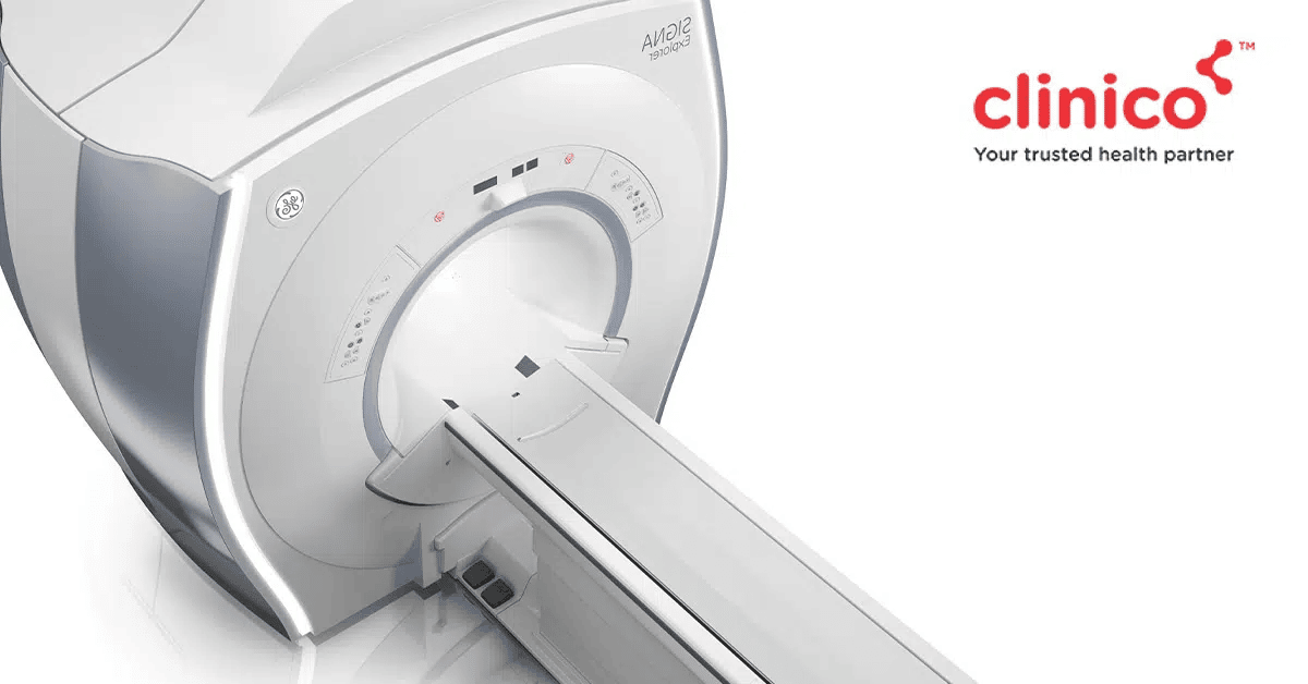
What is MRI Scan?
MRI or Magnetic Resonance Imaging is a non-invasive imaging technology that generates 3D detailed anatomical images of the organs & tissues in the human body. It makes use of a magnetic field and computer-produced radio waves to create the 3D images, which can then be viewed from a variety of angles.
Types of MRI Scans
Following are the 7 types of MRI scans based on the different indications:
- MRI Brain, Spine
- MRI Abdomen, Pelvis
- MRCP
- MR Urography
- MR Spectroscopy
- MRI Angiography
- MRI Venography
What is the purpose of an MRI Scan?
An MRI scan enables healthcare professionals to view the internal structures of the human body and diagnose & monitor a wide range of health conditions.
What is the diagnosis?
Through its detailed 3D images, MRI helps diagnose diseases impacting the muscles, organs, or other types of tissues in the human body. It also helps avoid/recognise the necessity of surgery for treatment and is especially beneficial for detecting health conditions related to the brain & spinal cord.
Following are the various health conditions an MRI scan can help diagnose:
- Brain & spinal cord conditions like stroke, multiple sclerosis, brain aneurysms, tumours & injuries
- Tumours or abnormalities in organs such as the liver, spleen, pancreas, reproductive organs, kidneys, bile ducts, bladder, heart, bowel & adrenal glands
- Heart & blood vessel structure problems like the abnormal size of aortic chambers, damage from a heart attack or heart ailment, inflammation, blockages, congenital heart disease, aneurysms & other heart issues
- Inflammatory bowel diseases such as Crohn’s disease or ulcerative colitis
- Joint & bone irregularities, tumours, abnormalities and infections
- Liver diseases such as cirrhosis
- Breast cancer
A special kind of MRI known as functional magnetic resonance imaging (fMRI) can be used to assess brain activity and view brain structure & blood flow to the brain. It’s particularly effective in detecting brain damage from a head injury, brain tumour, stroke, or the detrimental effects of diseases like Alzheimer’s.
What to Expect Before, During & After an MRI Scan?
Before the scan, you will be enquired regarding the following things that may interfere with the scan and alter the images:
- Metal implants of any kind
- Heart pacemakers or artificial heart valves
- Bullet fragments
- Insulin pumps or ongoing chemotherapy
If you have any anxiety disorders or claustrophobia, then you will be provided with a mild sedative to help decrease the uneasy feeling during the scan.
Once the scan begins, you will be asked to remain still, and you will hear constant loud humming noises & electronic beeping. The operator will be in communication with you via a radio. If you experience any sort of discomfort, you can alert the operator.
After the conclusion of the scan, there will be no recovery period as such, and you can straightaway get back to your daily routine.
What does a Brain MRI show?
A brain MRI generates 3D images of the brain from various angles. It allows healthcare professionals to view soft tissues inside the brain and detect any type of abnormality or anomaly such as:
- Blood clots or enlargement of blood vessels
- Tumours or cysts
- Brain tissue enlargement
- Swelling and inflammation
- Injuries or infections
- Developmental and structural irregularities
- Haemorrhage
- Stroke
- Fluid leaks
Conclusion
Overall speaking, an MRI Scan is a common yet crucial diagnostic imaging scan that is used worldwide to generate detailed, cross-sectional images of various human body parts for diagnosing a wide array of health conditions.
If you have been prescribed an MRI by your doctor, then get in touch with us at Clinico! We are one of Mumbai’s highly rated diagnostic chains offering world-class diagnostic services at our centres in Mulund, Bhandup & Thane.
Schedule your MRI Scan by contacting us 24×7 on 9504555555.
Hussain, Sarwat, et al. “MR urography.” Magnetic Resonance Imaging Clinics of North America 5.1 (1997): 95-106.
https://europepmc.org/article/med/8995127
Duijn, Jeffrey H., B. Matson, and Andrew A. Maudsley. “MR Spectroscopy ‘.” Radiology 183 (1992): 718.
https://www.lfmi.ninds.nih.gov/pubpdf/DuijnRadiology%201992.pdf
Poncelet, B., D. Baleriiaux, and C. Segebarth. “MRI angiography.” Recent advances in international radiology and new vascular imaging. 1989.
https://inis.iaea.org/search/search.aspx?orig_q=RN:20084937
