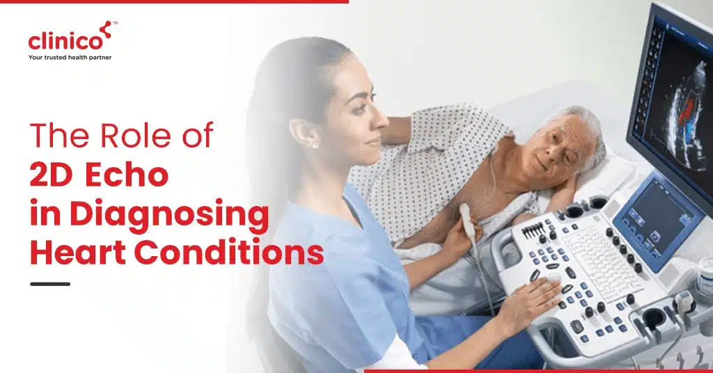
The heart, a vital organ in the human body, requires meticulous care and monitoring. Among the plethora of diagnostic tools available, the 2D Echocardiogram (2D Echo) stands out as a cornerstone in cardiovascular medicine. This non-invasive ultrasound test has revolutionized how cardiologists diagnose and manage heart conditions, offering a window into the heart’s intricate workings without the need for surgery. This blog delves into the significance of 2D Echo, highlighting its indispensable role in identifying a wide array of heart diseases.
What is a 2D Echocardiogram?
A 2D Echocardiogram is a type of ultrasound test that uses sound waves to produce images of the heart’s internal structures. Unlike traditional X-rays, it doesn’t expose patients to radiation, making it a safer alternative for ongoing monitoring. The images obtained can reveal vital information about the heart’s size, structure, and the functionality of its chambers and valves, providing a comprehensive view of its health.
The Diagnostic Power of 2D Echo
Identifying Valve Diseases
One of the primary applications of the 2D Echo is in the diagnosis of valve diseases. Valve issues can lead to significant health problems, including heart failure, if not addressed promptly. The detailed images produced by the 2D Echo allow doctors to see how the valves open and close with each heartbeat, identifying abnormalities such as stenosis (narrowing of the valve) or regurgitation (leakage of the valve).
Assessing Heart Chamber Size and Function
The size and functioning of the heart’s chambers are critical indicators of cardiovascular health. The 2D Echo can measure the dimensions of these chambers, detect abnormalities in their structure, and assess how well they pump blood. This is crucial for diagnosing conditions like cardiomyopathy, where the heart’s ability to pump blood is compromised.
Evaluating Heart Muscle Function
Heart muscle function is another area where the 2D Echo provides invaluable insights. The test can identify areas of the heart muscle that are not contracting well due to inadequate blood flow or damage from a previous heart attack. This information is crucial for determining the best course of treatment, which may include medications, lifestyle changes, or surgical interventions.
Advantages of 2D Echo
The 2D Echocardiogram offers several advantages over other diagnostic tests:
Non-invasive:
No needles, dyes, or radiation are involved, making it safe for repeated use.
Detailed imaging:
Provides a detailed view of the heart’s structure and function.
Real-time insights:
Allows for the immediate assessment of the heart.
Accessibility:
It can be performed in various settings, including hospitals, clinics, and even at the patient’s bedside.
Timely Interventions for Optimal Health Outcomes
The early detection of heart conditions through 2D Echo enables timely interventions, which can be pivotal in preventing the progression of heart disease. Whether it’s through medication, lifestyle changes, or surgery, early treatment based on 2D Echo findings can lead to better health outcomes and improve the quality of life for individuals with heart conditions.
Conclusion
The 2D Echocardiogram has cemented its place as an essential tool in the diagnosis and management of heart conditions. By offering a detailed, real-time look at the heart’s structure and function, it enables cardiologists to make accurate diagnoses and tailor treatments to individual patient needs. As medical technology advances, the role of the 2D Echo in ensuring optimal heart
outcomes continue to grow, highlighting its importance in the field of cardiovascular medicine.
FAQs
Q: Is the 2D Echocardiogram painful?
A: No, the 2D Echo is a painless procedure. Although the pressure of the transducer on your chest may cause slight discomfort, there is no pain involved.
Q: How long does a 2D Echo test take?
A:Typically, the test lasts about 30 to 60 minutes, depending on the complexity of the images needed.
Q: Do I need to prepare for a 2D Echo?
A: Generally, no special preparation is needed. You might be asked to wear comfortable clothing that provides easy access to your chest area.
Q: Can a 2D Echo detect all heart diseases?
A: While a 2D Echo is incredibly useful, it may not detect every heart condition. Your cardiologist might recommend additional tests based on your symptoms and health history.
Q: How often should I have a 2D Echo?
A: The frequency depends on your specific heart condition and risk factors. Your doctor will recommend a schedule based on your individual needs.

