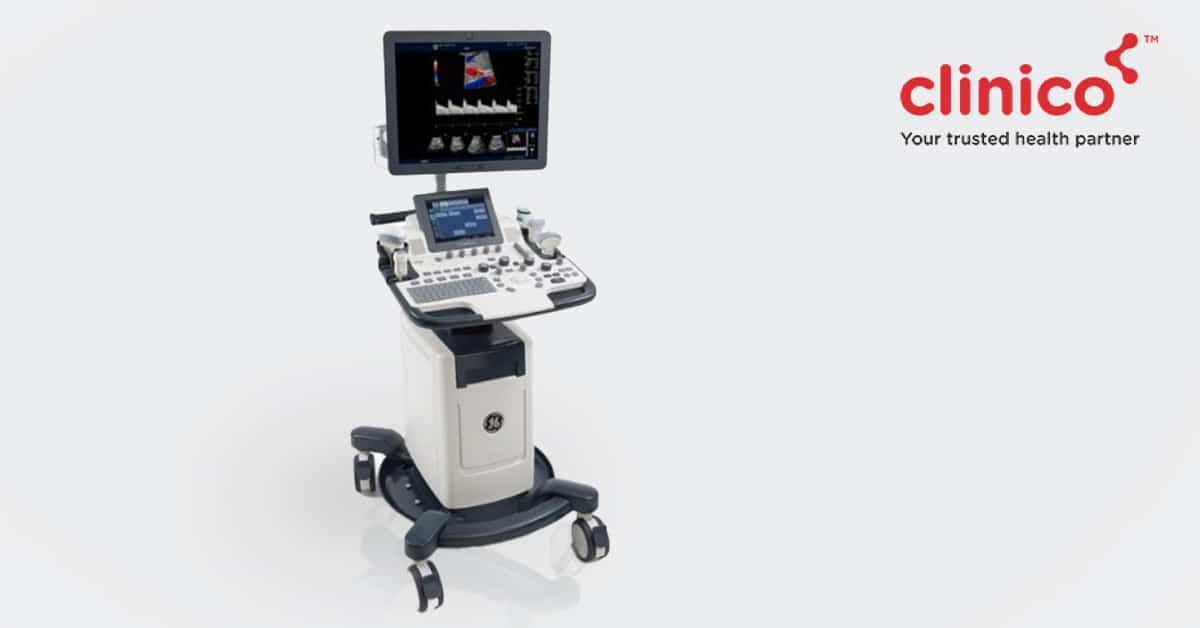
What is a 2D Echo Test?
2D Echo stands for 2-Dimensional Echocardiography. It’s a non-invasive imaging technique that helps assess the functioning of the heart and its various sections. This test generates images of the different parts of the heart using sound vibrations, making it simple for doctors to inspect for blockages, blood flow rate & damage.
What are the types of 2D Echo tests?
Many types of 2D Echo tests are in existence, with Transthoracic Echocardiography (TTE) the most common type employed in most tests. Other prominent types comprise Stress Echocardiography and Transesophageal Echocardiography.
What does the 2D Echo test result show?
The result of a 2D Echo test helps detect:
- Underlying heart diseases or issues
- Congenital heart diseases & blood clots/tumours
- Heart valve malfunction
- Abnormal blood flow inside the heart
What all things should be there in the results?
The 2D Echo test result includes information regarding the functioning of the heart, any malfunction, abnormal anatomy or atypical movement of heart structures.
With the help of this information, the doctor will diagnose the issue and decide on the course of treatment.
Follow up instructions after getting tested
The 2D Echo test doesn’t have any side effects, and no change in daily activities is usually required.
Conclusion
2D Echo is one of the most well-known and universally used tests for diagnosing heart diseases. It provides vital information regarding the cardiovascular health of a person.
Are you looking to get a 2D Echo test done for you or your loved ones?
Clinico is here to help!
We provide 2D Echo test service at our world-class diagnostic centres in Mulund, with state-of-the-art infrastructure and fast & accurate reports.
Schedule Your 2D Echo Test Today! Contact us 24×7 on 9504555555.
FAQ’s
A normal 2D echo test result mentions the absence of any heart malfunction and abnormal anatomy or atypical movement of heart structures.
The 60% in a 2D echo test report is the ejection fraction (percentage of blood pumped out of the filled left ventricle with each contraction of the heart). A normal ejection fraction ranges between 50-70%. – drop this question it depends on patient clinical condition too
Cardiovascular disease, heart valve disease, heart walls thickening, pericardial effusion & atrial fibrillation are the 5 abnormalities that can be detected in an echocardiogram.
“Cunha, C. L., et al. “”Echophonocardiographic findings in patients with prosthetic heart valve malfunction.”” Mayo Clinic Proceedings. Vol. 55. No. 4. 1980.”
https://europepmc.org/article/med/7359950


